You can look at your dog’s teeth and see perfectly white enamel. You might even think their smile is pristine. But that visual check misses up to 60% of the tooth structure. The roots, the bone support, and the hidden infections are invisible to the naked eye. This is why dog dental x-rays are not just an optional add-on; they are the gold standard in veterinary diagnostics.
Canine dental radiography allows veterinarians to see what is happening beneath the gum line. It reveals periodontal disease, abscesses, broken roots, and tumors long before they cause visible pain or swelling. Without these images, a vet is essentially guessing. With them, you get a precise map of your pet’s oral health.
The Hidden Half: Why Visual Exams Fail
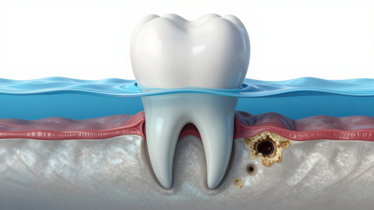
Think of a tooth like an iceberg. The part you see above the waterline is only a fraction of the total mass. In dogs, the clinical crown-the part visible in the mouth-is often less than half of the entire tooth structure. The rest is submerged in the gums and anchored by the alveolar bone.
When plaque builds up, it doesn't stop at the gum line. It creeps down. Bacteria invade the periodontal ligament and eat away at the bone that holds the tooth in place. By the time you notice redness, bad breath, or loose teeth, the damage is already severe. A visual exam can detect tartar on the surface, but it cannot measure bone loss. Only canine dental radiographs can quantify exactly how much supporting bone has been destroyed.
- Surface Level: Tartar buildup and gingivitis (red gums).
- Sub-Gum Level: Bone loss, root resorption, and hidden fractures.
- Deep Tissue: Abscesses, cysts, and neoplasms (tumors).
Without imaging, a vet might clean the top of the teeth while missing a massive infection at the root. That infection can spread to the heart, liver, and kidneys. Seeing the whole picture prevents systemic disease.
What Exactly Do Dental Radiographs Show?
Dental x-rays provide a two-dimensional image of three-dimensional structures. They reveal density differences. Dense objects like enamel and bone appear white (radiopaque). Less dense materials like air, soft tissue, and decayed areas appear dark (radiolucent). This contrast is what allows for diagnosis.
Here is a breakdown of specific conditions that radiographs uncover:
- Periodontal Disease Stages: Healthy bone looks sharp and defined around the tooth root. Diseased bone appears fuzzy or eroded. Vets use this to stage the disease from mild gingivitis to advanced periodontitis.
- Root Fractures: Dogs chew on hard objects like antlers, rocks, or nylon bones. These can crack the tooth root deep inside the jaw. A crack lets bacteria in, causing pain. You won't see this until the tooth falls out or an abscess bursts.
- Resorptive Lesions: Also known as Tooth Resorption Lesions (TRL) or FORL (Feline Odontoclastic Resorptive Lesions), these are painful spots where the body starts eating its own tooth structure. They look like dark holes in the tooth on an x-ray.
- Abscesses: An infection at the tip of the root creates a dark halo around the root end. This indicates pus accumulation and bone destruction.
- Retained Baby Teeth: If a puppy keeps both baby and adult teeth, the baby tooth can push against the adult one, displacing it. X-rays show if the baby root is dissolving naturally or needs extraction.
| Method | Visibility Depth | Accuracy for Bone Loss | Pain Detection |
|---|---|---|---|
| Visual Exam | Crown only | Low | None |
| Probing (Physical) | Gum pockets | Moderate | Low |
| Dental Radiographs | Full root & bone | High | High (via pathology) |
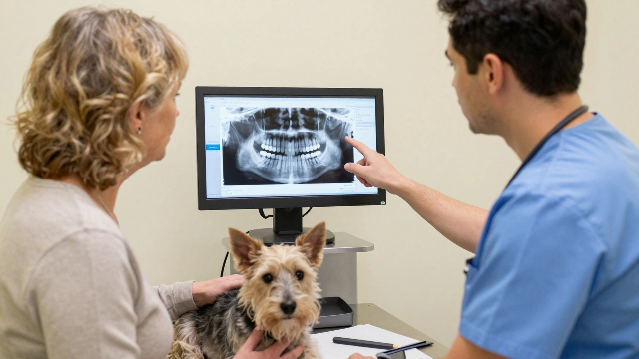
The Procedure: How Dental X-Rays Are Taken
You cannot take a high-quality dental x-ray of a conscious dog. Dogs move, breathe, and swallow. Motion blur makes the images useless for diagnosis. Therefore, veterinary dental radiography requires general anesthesia.
This is often the biggest concern for owners. "Why do I need to put my healthy dog under anesthesia just for pictures?" The answer is safety and quality. The process follows strict protocols to minimize risk:
- Pre-Anesthetic Blood Work: Checks kidney and liver function to ensure the dog can process anesthesia safely.
- Intubation: A tube is placed in the trachea to protect the lungs from water and debris during cleaning.
- Stabilization: The dog is monitored continuously (heart rate, oxygen, blood pressure) throughout the procedure.
- Positioning: The vet uses specialized sensors or film packets. For intraoral shots, the sensor goes inside the mouth next to the tooth. For extraoral shots, it goes outside the jaw.
Intraoral radiographs are the most common. They provide the highest detail because the sensor is close to the tooth. The vet takes multiple angles-usually mesial-distal and buccal-lingual-to create a complete view of each quadrant of the mouth. A full set of dental x-rays typically involves 18 to 24 individual images, depending on the size of the dog.
Risks vs. Benefits: Is It Worth It?
The risk of anesthesia in a healthy, young dog is extremely low, especially with modern monitoring equipment. The American Veterinary Medical Association (AVMA) considers dental radiographs a critical component of comprehensive veterinary care.
The benefits far outweigh the risks. Consider this scenario: A dog comes in for a cleaning. The vet sees a small hole in a premolar. Visually, it looks minor. But an x-ray reveals the root is fractured horizontally. If the vet leaves that tooth, the dog will develop a chronic abscess within weeks. That abscess can lead to sepsis. Extracting the tooth now saves the dog from future suffering and expensive emergency surgery.
Furthermore, radiographs help avoid unnecessary extractions. Sometimes a tooth looks loose due to inflammation, but the bone support is actually intact. With x-rays, the vet can save the tooth with proper cleaning and medication instead of pulling it prematurely.
Frequency and Age Guidelines
How often should your dog get dental x-rays? There is no one-size-fits-all rule, but guidelines exist based on age and risk factors.
Puppies: X-rays are crucial for puppies with retained deciduous (baby) teeth. If a baby tooth isn't falling out on its own, x-rays determine if the permanent tooth is impacted or if the baby root needs removal.
Adult Dogs (1-7 years): Annual exams are recommended. If the dog has good oral health, x-rays might be taken every 1-2 years. However, any change in behavior (drooling, dropping food, pawing at the face) warrants immediate imaging.
Senior Dogs (7+ years): Senior dogs are prone to periodontal disease and resorptive lesions. Annual dental x-rays are strongly advised. Early detection of bone loss can slow progression and preserve more teeth.
Breeds matter too. Small breeds like Chihuahuas, Pomeranians, and Yorkies are genetically predisposed to crowded teeth and rapid periodontal disease. They almost always require annual radiographs. Large breeds like Great Danes may have fewer issues but are prone to specific conditions like caudal enthesophytes (bone spurs) that x-rays detect.
Interpreting the Results: What Owners Should Know
After the procedure, your vet will discuss the findings. Don't be afraid to ask questions. Here are key terms you might hear:
- Alveolar Crest: The top edge of the bone holding the tooth. If it's lower than normal, bone loss has occurred.
- Periodontal Ligament Space: The thin black line between the tooth root and the bone. If it widens, it suggests infection or trauma.
- Periapical Radiolucency: A dark area at the root tip. This usually means an abscess.
- Internal Resorption: Darkening inside the tooth chamber, indicating the body is breaking down the tooth from the inside.
Your vet will likely present a treatment plan. Options include:
- Extraction: Removing the tooth entirely. This is the most common solution for severely diseased teeth.
- Root Canal Therapy: Saving the tooth by removing the infected nerve. This is rare in dogs but possible for large, important teeth.
- Hemisection: Splitting a multi-rooted tooth and removing only the damaged half.
- Monitoring: If the issue is minor, the vet might recommend re-evaluating in 6 months.
Cost Considerations
Dental x-rays add to the cost of a dental cleaning. Prices vary by location and clinic type. In urban areas like Portland, Oregon, a full dental prophylaxis with x-rays can range from $300 to $800. Rural clinics may charge less. Specialty hospitals may charge more for advanced imaging techniques like digital panoramic x-rays.
However, consider the cost of neglect. Treating a ruptured abscess, facial swelling, or systemic infection can cost thousands. Preventative radiographs are an investment in long-term health. Many pet insurance plans now cover diagnostic imaging if pre-existing conditions are ruled out. Check your policy.
Are dental x-rays safe for dogs?
Yes, dental x-rays are very safe. The radiation dose used in veterinary dental radiography is extremely low, comparable to a few minutes of natural background radiation. The sensors are shielded, and the exposure time is fractions of a second. The risk from radiation is negligible compared to the benefit of accurate diagnosis.
Can my dog get dental x-rays without anesthesia?
No, not for a comprehensive exam. High-quality diagnostic images require the dog to remain completely still. Conscious sedation is not sufficient for the precision needed. Attempting to x-ray a moving animal results in blurry images that cannot diagnose bone loss or fractures accurately. General anesthesia ensures both safety and diagnostic quality.
How often should senior dogs have dental x-rays?
Senior dogs (age 7 and older) should ideally have dental x-rays annually. As dogs age, they are more susceptible to periodontal disease, tooth resorption, and oral tumors. Annual imaging allows vets to catch these issues early when treatment is less invasive and more effective.
What does "bone loss" mean on a dental x-ray?
Bone loss refers to the destruction of the alveolar bone that supports the teeth. On an x-ray, healthy bone appears dense and white. Diseased bone appears darker and less defined. Mild bone loss affects the top edge of the bone. Severe bone loss extends down the root, potentially leading to tooth mobility and loss.
Do all dogs need dental x-rays?
While not legally required, professional veterinary organizations strongly recommend dental x-rays for all dogs undergoing anesthesia for dental cleaning. Since 60% of the tooth is hidden below the gum line, skipping x-rays means ignoring the majority of potential problems. Even dogs with perfect-looking teeth can have significant underlying disease.
What is the difference between intraoral and extraoral x-rays?
Intraoral x-rays are taken with the sensor inside the mouth, next to the specific tooth. They provide high-detail images of individual teeth and surrounding bone. Extraoral x-rays are taken with the sensor outside the jaw. They show broader views of the jaw structure but lack the fine detail needed for diagnosing periodontal disease or small fractures.
Can dental x-rays detect oral cancer?
Yes, dental x-rays can detect signs of oral tumors. Tumors often destroy bone, creating irregular dark areas on the radiograph. While x-rays suggest the presence of a mass, a biopsy is required for a definitive cancer diagnosis. Early detection via x-rays improves prognosis significantly.
Why did my vet recommend extracting a tooth that looked fine?
The tooth likely had hidden damage revealed by x-rays. Common reasons include root fractures, severe bone loss, internal resorption, or abscesses. Leaving a compromised tooth causes chronic pain and infection. Extraction removes the source of pain and prevents systemic health issues.